

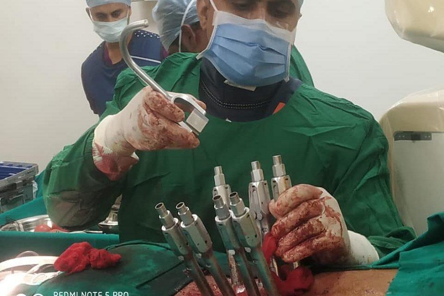
When it comes to complex spine procedures, precision is critical. Spine surgeons must navigate a network of delicate structures, nerves, discs, blood vessels, and vertebrae, all in proximity. A slight miscalculation can lead to serious consequences, including nerve damage or ineffective treatment. That’s why imaging has always been an integral part of spinal surgery. In recent years, real-time imaging has emerged as a valuable tool, giving surgeons the ability to see inside the body with remarkable clarity during procedures. Dr. Larry Davidson, a leader in spinal surgery, recognizes that the integration of this technology has played a major role in improving outcomes and reducing complications.
Real-time imaging refers to technologies such as fluoroscopy, intraoperative CT, and neuronavigation that allow spine surgeons to visualize anatomical structures in motion during surgery. Unlike preoperative scans, which provide a static view, these tools give dynamic feedback, enhancing precision and control. Surgeons can see how instruments interact with tissue, verify the placement of implants, and adjust on the fly, an advantage especially important in minimally invasive procedures where the surgical field is limited.
The Shift Toward Real-Time Guidance
Before the widespread use of real-time imaging, many spinal procedures relied heavily on preoperative planning and the surgeon’s anatomical knowledge. While effective, this approach left little room for intraoperative adjustments. With real-time imaging, procedures become more adaptable. Surgeons can confirm alignment, monitor movement, and track changes throughout the operation.
Fluoroscopy remains a widely used modality for real-time imaging, providing continuous X-ray guidance during instrumentation and implant placement. Its portability and accessibility make it a staple in many spine operating rooms. Surgeons can visualize needles, guidewires, and cement in procedures like kyphoplasty and vertebroplasty, reducing the risk of misplacement or leakage.
Read More: Evan Bass Men’s Clinic Discusses the Impact of Fatherhood on Personal Development
Intraoperative CT and neuronavigation systems take it a step further. These technologies offer three-dimensional, high-resolution images that are updated throughout the procedure. When used in conjunction with navigation platforms, they help create a “GPS-like” system that allows for precise trajectory planning and tool guidance. These advances have not only improved surgical accuracy but also decreased the likelihood of revision surgeries.
Enhancing Safety in Cement-Based Procedures
Real-time imaging plays a pivotal role in cement augmentation procedures, such as vertebroplasty and kyphoplasty, which are frequently used to treat spinal compression fractures. In these surgeries, cement is injected into weakened or collapsed vertebrae to provide support and relieve pain. But the process must be handled with care. Improper placement or leakage of cement can lead to serious complications, including nerve compression or pulmonary embolism.
Imaging helps prevent these outcomes by allowing the surgeon to monitor cement flow in real time. If the cement begins to migrate toward sensitive areas, the injection can be paused or redirected. This immediate feedback helps surgeons intervene before problems occur, improving patient safety.
As these procedures become more common in older populations and those with bone density issues, the value of imaging continues to increase. Being able to deliver precise amounts of cement to the exact location needed without affecting nearby structures contributes to both efficacy and peace of mind for patients and providers alike.
Supporting Minimally Invasive Spine Surgery
Real-time imaging is especially vital in Minimally Invasive Spine Surgery (MISS). These procedures, which rely on small incisions and specialized tools, offer a number of benefits: reduced pain, shorter hospital stays, and faster recovery. But they also present challenges due to the limited visibility inside the body.
Read More: Best Health Insurance Plans in India 2025: Comparison Guide
Here, imaging functions as the surgeon’s eyes. It compensates for the restricted access by providing clear and continuous visualization of the surgical site. Tools can be introduced through narrow channels and guided precisely to the target without damaging surrounding tissue. Implants, screws, and grafts can be positioned with millimeter-level accuracy.
As minimally invasive techniques expand to treat a broader range of spinal conditions, from herniated discs to spinal stenosis, real-time imaging has become a critical enabler. It helps ensure that these procedures remain as safe and effective as possible, even in anatomically complex or high-risk cases.
Educating and Empowering the Surgical Team
One often overlooked benefit of real-time imaging is its impact on surgical education and teamwork. The visual feedback allows all members of the operating team to see what the surgeon sees, improving communication and coordination. Trainees gain valuable insights by observing how decisions are made based on live images, while anesthesiologists and nurses can better anticipate the next steps.
This shared view also supports documentation and postoperative analysis. Surgeons can review images to evaluate the procedure’s success, identify potential issues, and refine their technique for future cases. For teaching hospitals and research centers, this data is a valuable resource for improving surgical outcomes and developing best practices.
Patient Benefits and Expectations
Patients undergoing spinal surgery today often arrive more informed than ever. Many have researched treatment options and are aware of the differences between traditional and minimally invasive approaches. Real-time imaging is one feature that helps set expectations for a more precise and less disruptive experience.
Real-time imaging aligns with patients’ growing desire for personalized, high-quality care by enabling safer procedures, reducing complications, and contributing to better outcomes. In cases involving spinal fractures or instability, the ability to visualize and address issues during the surgery itself can mean less postoperative discomfort and a quicker return to daily activities.
Dr. Larry Davidson notes, “As physicians, we are here to treat people, not just conditions. The technology is only as effective as the thoughtfulness we bring to its use.” This perspective reflects the integration of compassion and innovation that defines modern spine surgery. It’s a reminder that while tools may evolve, the human connection remains at the heart of healing.
The Path Forward
As more facilities adopt real-time imaging systems, the standard of care for spine surgery continues to shift. While not every case requires the most advanced tools, the ability to tailor imaging approaches to the patient’s condition and the surgeon’s strategy is a major advantage. Whether it’s ensuring precise screw placement in a fusion procedure or guiding cement flow during kyphoplasty, the benefits of real-time feedback are hard to ignore.
Future developments in this space are expected to focus on improving image resolution while reducing radiation exposure and integrating artificial intelligence to assist with interpretation. For now, real-time imaging hasreshaped what is possible in spine surgery, offering a clearer path to better outcomes and safer care for those living with back pain or spinal disorders.